Have you ever bent over and suddenly been unable to stand up straight from lower back pain? Have you ever woken up, rolled over and thought “ouch” this isn’t going to be a very good day, as the pain radiates from your lower back down your leg to behind your knee? Lower back pain affects our quality of life and our job performance. It affects your horse’s life and performance as well.
We are able to share our symptoms of lower back pain with our family, friends and medical professionals. How do our horses share their symptoms? If we listen, they are trying to tell us. Some of the outwards signs would include: lack of drive from their hindquarters, hind end lameness that is hard to diagnose, tenderness to the touch over the rump, and not wanting to round or lift the back while being worked. If your horse has some of these symptoms, what can you do to diagnose and treat them? First understand how horses’ backs function.
Anatomy/Physiology
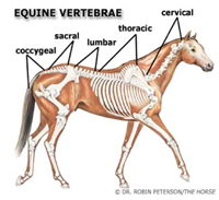
Figure 1. Drawing of the equine vertebral column within the horse’s body. Reprinted with permission from the illustrator Dr. Robin Peterson.
The horse’s vertebral column is made up of approximately 54 vertebrae that surround and protect the horse’s spinal cord (Fig.1). Starting at the poll area of a horse there are 7 cervical (neck) vertebrae and 18 thoracic (chest) vertebrae. Each thoracic vertebra has a rib attachment. In the lower back, most horse have 6 lumbar vertebrae, except for some “short backed” Arabians that only have 5 lumbar vertebrae. The sacrum is made up of 5 fused vertebrae. The last set of coccygeal (tail) vertebrae can range in number from 5-18 depending on the breed and tail length of the horse. The sacrum and the ilium (part of the pelvis) are attached together at the sacroiliac joint (Fig 2.). This joint is the link between the hind limb extremities and the spine in a horse. The sacroiliac joint sustains high loading force during athletic activity and can be the source of low back pain in horses.
Symptoms
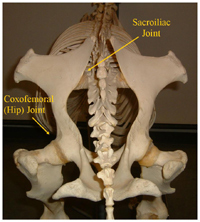 Figure 2. Picture of an equine skeletal model from behind showing the pelvis, sacroiliac joint, and the coxofemoral (hip) joint.
Signs of sacroiliac pain in horse may include poor performance or lack of hind end impulsion. Horses with this problem also have had mild, chronic hind end lameness that is difficult to diagnose and does not resolve with traditional lower leg joint blocks or therapy. In a necropsy survey of 36 Thoroughbred racehorses, all specimens had various degrees of arthritis in their sacroiliac joint.1 In fact 92% of the specimens had moderate to severe degenerative changes in the pelvic area. To determine if the horse has a problem in the pelvis or sacroiliac region, a thorough physical and soundness exam should be performed by a veterinarian. The lameness exam should be started at the lower leg, proceed with joint flexions, diagnostic nerve or joint blocks, radiographs, ultrasound scan, and possibly nuclear bone scintigraphy. If a pelvic problem is suspected, one must determine if it is the primary cause of lameness or if the pain secondary to another sore area. More often than not, sore muscles or “trigger points” in the sacroiliac region are due to compensation of pain elsewhere in the body and are not a “true” pelvic problem. Keeping this in mind, a comprehensive lameness diagnostic exam is crucial.
Diagnosis
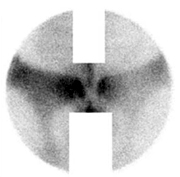 Figure 3. Nuclear Bone Scintigraphy scan of a horse with increased rate of absorption of the isotope in the left sacroiliac joint.
Nuclear bone scintigraphy is the best way to diagnose pelvic problems. This is a non-invasive technique in which the horse is given an intravenous injection of a radioactive isotope. The whole horse is scanned to see where the isotope is taken up by the horse’s skeleton (Fig. 3). The most painful areas have in increased rate of absorption of the radioactive isotope and can be detected by the scan.2 This procedure has a high percentage of detecting a pelvic problem if there is one.
The sacroiliac region also involves several ligaments and soft tissue structures. An ultrasound scan of the area may detect bone and ligament injuries. This technique is also non-invasive. It requires high level ultrasound skills, high quality machine and probe, and knowledge of the pelvic bone and soft tissue anatomy. The veterinarian should scan the external pelvic structures and also perform an internal rectal ultrasound exam.
Radiographs of the pelvis may also be performed depending on the size of the horse. In some smaller or young horses, with less muscle, the coxofemoral (hip) joint can be x-rayed standing. However most pelvis or hip radiographs must be taken under general anesthesia so the horse can be laid on its back. One must always keep in mind that there are always risks involved in putting a horse under or recovering from general anesthesia. These risks are increased in horses with hind end lameness.
Treatment Options
If a pelvic problem is diagnosed, there are some treatment options. Most pelvic problems are managed, however not always cured. A stress fracture of the pelvis as long as there is not major displacement will heal with time. Shockwave therapy can speed healing time. If the pelvis is completely fractured there is a poor prognosis. The sacroiliac area can be can be injected with corticosteroids, sarapin, or other counter-irritants. The joint itself is difficult to inject, however the medication around the area has been shown to provide some pain relief.
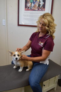
Chiropractic and acupuncture therapy can also help this area. When discussing equine chiropractic therapy you may have heard the term subluxation complex. This term means different things to different people. In traditional veterinary medical terms, subluxation is defined as a partial dislocation of a joint. This has a poor prognosis and requires medical and possibly surgical attention. A subluxation complex in chiropractic terms means that the joint surfaces are not in the correct position by millimeters. The joint itself still functions, but not as well. The subluxation complex may also cause impingement of the spinal nerve in that area and thus decrease nervous enervation to the surrounding area. There is loss of normal motion due to pain, muscle spasms, or joint stiffness. Chiropractic therapy involves examining the entire horse to locate subluxation complexes and tight muscle and painful areas. The subluxation complexes are adjusted by delivering a short, sharp thrust with the doctor’s hands or an instrument to a specific joint at a specific angle on the horse’s spine or limbs. One joint is worked on at a time. Many times the doctor must stand on a stool or bale to achieve the correct alignment angle. If the incorrect angle or thrust is applied, great damage can be done to the animal. It is important to make sure that the doctor you have work on your animal is properly trained and certified in animal chiropractic. The chiropractic therapy should be performed by a doctor certified in Animal Chiropractic through the American Veterinary Chiropractic Association. You can locate a qualified doctor in your area by searching the AVCA website at www.animalchiropractic.org. The goal of the chiropractic therapy is to restore normal range of motion in the horse’s vertebral column and limbs, release impinged nerves and thus muscle spasms, and alleviate pain in the animal.
Acupuncture and chiropractic therapy can be mutually enhancing and it is beneficial to integrate both modalities for treating pelvic problems in horses. Equine acupuncture therapy originates from Traditional Chinese Veterinary Medicine over 3000 years ago. The therapy involves stimulating a particular point on the body with a particular method to produce a therapeutic effect. Acupuncture points are located throughout the body on acupuncture meridians. There are several important and effective acupuncture points around the sacroiliac region of the horse. The overall goal of acupuncture is to stimulate internal anti-inflammatory and pain relief mechanisms to balance the body, facilitate tissue repair and prevent injury or disease. Integrating the various forms of therapy can decrease the possibilities of injuries and optimize the health and performance of our equine athletes.
Recognizing, diagnosing, and treating your horses’ lower back pain is critical to their comfort and to their success as a performance horse.
References:
1. Haussler, KK. Functional Anatomy and Pathophysiology of Sacroiliac Joint Disease. AAEP Proceedings 2004; 50:361-366.
2. Dyson, SJ, Murray, RC, Branch M. Uses and Limitations of Nuclear Scintigraphy for Evaluation of the Sacroiliac Region. AAEP Proceedings 2004; 50:379-384.